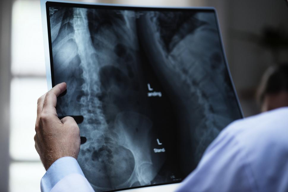
Planning for spine surgery begins well before the patient enters the operating room. One tool in this process is diagnostic imaging. While clinical symptoms and physical exams offer valuable insight, imaging creates a detailed map of the spinal anatomy. With precise visual data, physicians can make better decisions about whether a surgical approach is needed and what kind of procedure will offer the best results.
MRI and CT scans often play a key role in evaluating structural changes, spinal cord compression, and nerve root involvement. The choice between these options depends on what the spine surgeon needs to examine. MRI is often used for soft tissues, while CT scans give more clarity on bone structure.
MRI and CT Scans
Magnetic resonance imaging shows inflammation, disc damage, and spinal cord involvement with excellent clarity. It allows providers to evaluate disc herniation, degenerative conditions, and ligament injury without using radiation. This makes it one of the most frequently used tools in spine surgery planning, especially when nerve symptoms are present. CT scans, on the other hand, provide precise views of bones and joints. When hardware placement is expected or spinal fractures are suspected, these scans help define the anatomy in three dimensions. Sometimes, both MRI and CT are needed to form a complete picture.
Real-Time Data
Intraoperative imaging has transformed how many spine surgeons operate. Fluoroscopy and navigation systems deliver real-time images during the procedure. This level of accuracy helps guide hardware placement, align structures correctly, and reduce surgical risks. These technologies contribute to shorter operation times and fewer complications. They also give the surgical team immediate feedback, which improves confidence in the chosen approach. While not every procedure requires navigation, it is becoming more common in complex or minimally invasive cases.
Planning Complex Procedures
Spine surgery can involve delicate structures and narrow margins for error. Imaging allows surgeons to plan the angle, depth, and hardware size in advance. For conditions like scoliosis, spinal instability, or tumor removal, this preparation is key to both safety and outcomes. 3D modeling based on patient-specific imaging can also be created in select cases. These models offer even more visualization and are used for planning multi-level fusions or corrective surgeries. When imaging guides each step, the surgical team can operate with greater precision.
Patient-Specific Approach
Every spine is unique, and imaging supports a tailored approach to treatment. By identifying anatomical differences, providers can adjust their methods to better match the patient’s condition. A strategy that works well for one person may not be right for another. Visual data supports those choices and improves communication between the provider and patient. Imaging can also reveal whether a patient may benefit from non-surgical treatments first. This allows for a full understanding of the issue before making a recommendation.
The role of imaging extends beyond the operating room. Postoperative scans help monitor healing, assess hardware position, and identify any early signs of complications. If patients continue to experience pain or numbness after the procedure, new imaging may help locate the cause.
These follow-up scans offer another chance to protect outcomes. If adjustments are needed, imaging guides next steps. It also reassures both the patient and provider that healing is progressing as expected.
Improving Spine Surgery Outcomes
The use of imaging in spine surgery is not only about planning—it’s also about long-term success. Each scan contributes to a clearer understanding of what the patient needs and how to reach that goal safely. With consistent use of advanced imaging, providers can improve alignment, reduce the chance of revision surgery, and support better function for the patient. Modern spine surgery relies heavily on this technology. As tools become more advanced, so does the potential for improved recovery and surgical accuracy.

