An echocardiogram is a medical test that uses ultrasound technology to create detailed images of the heart. For individuals with known cardiac conditions, regular echocardiograms can be a component of their long-term management plan. This consistent monitoring supports ongoing care coordination by providing a clear view of the heart’s status over time, helping specialists track changes and make informed decisions about patient care.
What Is an Echocardiogram?
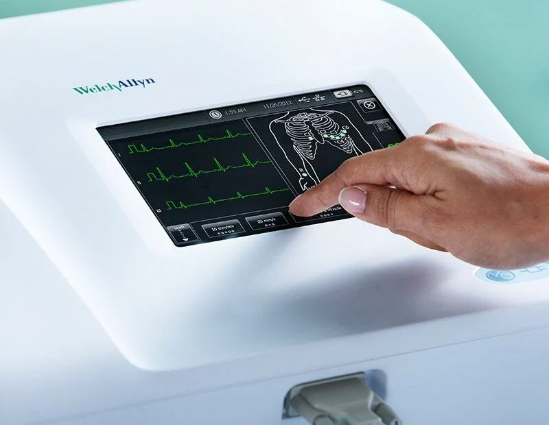
An echocardiogram is an imaging test that produces detailed images of the heart. A technician applies a gel to the chest and moves a transducer across the skin. The transducer emits high-frequency sound waves that travel through the body, bounce off the heart’s structures, and return to the probe.
This test can provide detailed information about the heart. It shows the size of the heart’s chambers and can reveal if they are enlarged. The imaging also allows for an assessment of the thickness and motion of the heart walls, which is useful for evaluating the heart muscle. The movement and function of the heart’s valves can be observed, showing how well they open and close with each heartbeat.
What Are the Different Types?
Several types of echocardiograms are available, each suited for different clinical situations. The standard transthoracic echocardiogram (TTE) is the most frequently performed version. It is noninvasive and provides a general overview of heart structure and function from outside the chest wall.
In some cases, a more detailed view is needed. A transesophageal echocardiogram (TEE) may be performed for this purpose. During a TEE, a specialized probe at the end of a thin, flexible tube is guided down the esophagus. Because the esophagus is located directly behind the heart, this method bypasses the ribs and lungs, often resulting in clearer, higher-resolution images of certain heart structures.
Another variation is the stress echocardiogram. This test is used to evaluate the heart’s performance under physical load. Images of the heart are taken both at rest and immediately after exercise. Comparing the resting and stress images can help a specialist assess how the heart responds to exertion.
What Conditions Do They Monitor?
Echocardiography is utilized to monitor a wide range of established heart conditions. It is often used to evaluate heart valve disorders, such as leaky valves or stenosis. It does this by tracking changes in their severity over time. For individuals with heart failure, periodic echocardiograms can provide insight into the heart’s pumping strength by observing trends. Following a heart attack, the test may be used to evaluate changes in heart muscle tissue and overall function. The procedure may offer information on structural changes to the heart that can be associated with certain heart rhythm disorders.
Meet With a Heart Specialist
An echocardiogram provides valuable visual information about your heart, but the results are just one part of a comprehensive cardiac evaluation. A qualified heart specialist can interpret these images in the context of your overall health, symptoms, and the results of other medical tests. Scheduling a consultation allows you to discuss your specific situation and determine if an echocardiogram is appropriate for you.